Female Normal Pelvic Ultrasound Report - Dr Naeem Taj Taj Surgery Offical Page Ultrasound Report Of Left Sided Gall Bladder Facebook - It involves an ultrasound scan as well as a biochemical test to assess the risk of ovarian cancer.
byAdmin•
0
Female Normal Pelvic Ultrasound Report - Dr Naeem Taj Taj Surgery Offical Page Ultrasound Report Of Left Sided Gall Bladder Facebook - It involves an ultrasound scan as well as a biochemical test to assess the risk of ovarian cancer.. Evaluation for signs or symptoms of pelvic infection 13. Make sure to have a drape/sheet and cover the patient properly. Normal bilateral ovaries without evidence of. Further charac terization of a pelvic abnormality noted on another imaging study 14. The sound waves create a picture on a video monitor.
Complete pelvic ultrasound (upeltv) this is a complete pelvic ultrasound exam, including transabdominal and transvaginal. If a male sonographer is doing the scan, there will need to be a female chaperone present for the transvaginal or translabial portion of the exam. Normal pelvic ultrasound nazari l, assistant professor ob/gyn shahid beheshti university of medical sciences. The endometrial stripe measures __ cm in diameter and is normal in echogenicity. The sound waves create a picture on a video monitor.
Http Www Arcxray Com Au Images Referrer Pdf Transvaginal 20ultrasound 20patient 20information Pdf from Make sure to have a drape/sheet and cover the patient properly. Evaluation of excessive bleeding, pain, or signs of infection after pelvic surgery, delivery, or abortion 16. Report whether any mass is delineated. Evaluation for signs or symptoms of pelvic infection 13. A pelvic ultrasound allows quick visualization of the female pelvic organs and structures including the uterus, cervix, vagina, fallopian tubes and ovaries. No cysts are identified (a __ cm round, anechoic cyst is noted or normal follicles are identified). Both ovaries should be identified and measured if appropriate again in 3 planes. Pelvic ultrasound is usually the initial modality for imaging gynecologic pathology, including acute pelvic pain and chronic pelvic pain.
A pelvic ultrasound is a noninvasive diagnostic exam that produces images that are used to assess organs and structures within the female pelvis.
Female pelvis ultrasound sonography plays the primary role in imaging of the female pelvis. Primary indications for female pelvic us examination are pelvic pain, abnormal vaginal bleeding, and suspicion of pelvic mass. An ultrasound of the female pelvis may be performed by examination of the abdomen, called transabdominal, or by using a special probe designed to be inserted into the vagina, called transvaginal. Ultrasound imaging uses soundwaves to create pictures of the inside of the body. However, it is considered more invasive than the transabdominal approach. • normal and ectopic pregnancy implantation can be accurately Within the right ovary, there is a 2.5 cm thick walled cyst with crenulated inner margin and peripheral vascularity consistent with a corpus luteum. A transabdominal (ta) evaluation and a transvaginal (tv) / endovaginal (ev) evaluation. Further charac terization of a pelvic abnormality noted on another imaging study 14. A pelvic ultrasound is a noninvasive diagnostic exam that produces images that are used to assess organs and structures within the female pelvis. Measurements should be recorded for simple/complex/dermoid cysts. Transvaginal ultrasound is a test used to look at a woman's uterus, ovaries, tubes, cervix and pelvic area. Both ovaries should be identified and measured if appropriate again in 3 planes.
Multiple sonographic images of the pelvis were assessed for gray scale appearance and color doppler flow. Evaluation for signs or symptoms of pelvic infection 13. Make sure to have a drape/sheet and cover the patient properly. The exam normally involves two components: The type of examination you will need will depend on what your referring doctor has requested and the nature of the clinical condition being investigated.
Noninvasive Ultrasound Diagnosis Of Endometriosis from cdn.sanity.io It allows your doctor to see your bladder, cervix, uterus, fallopian tubes, and ovaries. Multiple sonographic images of the pelvis were assessed for gray scale appearance and color doppler flow. It involves an ultrasound scan as well as a biochemical test to assess the risk of ovarian cancer. By dr attiya khan and mr rehan khan. Normal bilateral ovaries without evidence of. A transabdominal (ta) evaluation and a transvaginal (tv) / endovaginal (ev) evaluation. Primary indications for female pelvic us examination are pelvic pain, abnormal vaginal bleeding, and suspicion of pelvic mass. The ultrasound report needs to strike a balance between conciseness and sufficient clinical detail.
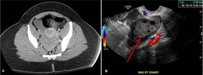
Within the right ovary, there is a 2.5 cm thick walled cyst with crenulated inner margin and peripheral vascularity consistent with a corpus luteum.
Transvaginal ultrasound is a test used to look at a woman's uterus, ovaries, tubes, cervix and pelvic area. An ultrasound of the female pelvis may be performed by examination of the abdomen, called transabdominal, or by using a special probe designed to be inserted into the vagina, called transvaginal. This gynaecological scan is available to post menopausal women (i.e. It is normal in echogenicity with normal vascular flow. Please refer to the articles on pelvic ultrasound, for a discussion about this study, and on normal genitourinary tract. 1 year after last period). Ultrasound imaging uses soundwaves to create pictures of the inside of the body. An ultrasound of the pelvis is typically used to look at the bladder, ovaries, uterus, cervix, and fallopian tubes (some of these are known as the female reproductive organs). A hand held device called a transducer (also called a probe or wand) sends and receives these soundwaves. Ultrasound of the female pelvis is important for diagnostic accuracy. The left ovary measures __ x __ x __ cm in diameter and demonstrates normal vascular flow and echogenicity. A pelvic ultrasound allows quick visualization of the female pelvic organs and structures including the uterus, cervix, vagina, fallopian tubes and ovaries. Normal bilateral ovaries without evidence of.
Multiple sonographic images of the pelvis were assessed for gray scale appearance and color doppler flow. Female pelvis ultrasound sonography plays the primary role in imaging of the female pelvis. A transvaginal ultrasound, also called an endovaginal ultrasound, is a type of pelvic ultrasound used by doctors to examine female reproductive organs. It is normal in echogenicity with normal vascular flow. A pelvic ultrasound is a test that uses sound waves to make a picture of the inside of the lower belly (pelvis).
Non Visualization Of The Ovary On Ct Or Ultrasound In The Ed Setting Utility Of Immediate Follow Up Imaging Springerlink from media.springernature.com The left ovary measures __ x __ x __ cm in diameter and demonstrates normal vascular flow and echogenicity. Fed up with deciphering jargon, dr attiya khan asked consultant gynaecologist mr rehan khan for a plain language guide to understanding pelvic ultrasounds. A pelvic ultrasound is a safe procedure that can be slightly uncomfortable. Ultrasound imaging uses soundwaves to create pictures of the inside of the body. A pelvic ultrasound is a test that uses sound waves to make a picture of the inside of the lower belly (pelvis). Ct and mr are supplemental techniques used when the us examination is equivocal and in the staging of pelvic malignancy. The test can be done in two ways: No cysts are identified (a __ cm round, anechoic cyst is noted or normal follicles are identified).
This includes the uterus, fallopian tubes,.
• a variety of gynecologic conditions (congenital uterine anomalies, endometrial abnormalities, submucosal fibroids, and adenomyosis) can be evaluated with 3d ultrasound and may obviate need for further imaging. If a male sonographer is doing the scan, there will need to be a female chaperone present for the transvaginal or translabial portion of the exam. Female pelvis ultrasound sonography plays the primary role in imaging of the female pelvis. Ultrasound of the female pelvis is important for diagnostic accuracy. The test is performed on men and women of all ages. Please refer to the articles on pelvic ultrasound, for a discussion about this study, and on normal genitourinary tract. 1 year after last period). This review presents the normal and pathologic development of the gonads and genitourinary tract and addresses the role of ultrasonography in the diagnosis and management of gynecologic disorders of the pediatric pelvis, including ambiguous genitalia, prepubertal bleeding, primary amenorrhea, pelvic mass, and pelvic pain. Other codes that can be used as needed are upelc, upell, utvag. A transvaginal ultrasound, also called an endovaginal ultrasound, is a type of pelvic ultrasound used by doctors to examine female reproductive organs. No cysts are identified (a __ cm round, anechoic cyst is noted or normal follicles are identified). This gynaecological scan is available to post menopausal women (i.e. A pelvic ultrasound is a noninvasive diagnostic exam that produces images that are used to assess organs and structures within the female pelvis.